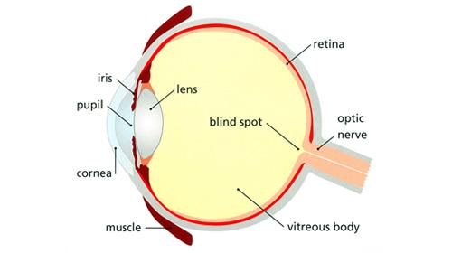
Anatomy of the eye
Our eyes might be small, but they provide us with what many people consider to be the most important of our senses – vision.
How vision works
Vision occurs when light enters the eye through the pupil. With help from other important structures in the eye, like the iris and cornea, the appropriate amount of light is directed towards the lens.
Just like a lens in a camera sends a message to produce a film, the lens in the eye 'refracts' (bends) incoming light onto the retina. The retina is made up by millions of specialised cells known as rods and cones, which work together to transform the image into electrical energy, which is sent to the optic disk on the retina and transferred via electrical impulses along the optic nerve to be processed by the brain.

Anatomy of the eye
What makes up an eye
- Iris: regulates the amount of light that enters your eye. It forms the coloured, visible part of your eye in front of the lens. Light enters through a central opening called the pupil.
- Pupil: the circular opening in the centre of the iris through which light passes into the lens of the eye. The iris controls widening and narrowing (dilation and constriction) of the pupil.
- Cornea: the transparent circular part of the front of the eyeball. It refracts the light entering the eye onto the lens, which then focuses it onto the retina. The cornea contains no blood vessels and is extremely sensitive to pain.
- Lens: a transparent structure situated behind your pupil. It is enclosed in a thin transparent capsule and helps to refract incoming light and focus it onto the retina. A cataract is when the lens becomes cloudy, and a cataract operation involves the replacement of the cloudy lens with an artificial plastic lens.
- Choroid: the middle layer of the eye between the retina and the sclera. It also contains a pigment that absorbs excess light so preventing blurring of vision.
- Ciliary body: the part of the eye that connects the choroid to the iris.
- Retina: a light sensitive layer that lines the interior of the eye. It is composed of light sensitive cells known as rods and cones. The human eye contains about 125 million rods, which are necessary for seeing in dim light. Cones, on the other hand, function best in bright light. There are between 6 and 7 million cones in the eye and they are essential for receiving a sharp accurate image and for distinguishing colours. The retina works much in the same way as film in a camera.
- Macula: a yellow spot on the retina at the back of the eye which surrounds the fovea.
- Fovea: forms a small indentation at the centre of the macula and is the area with the greatest concentration of cone cells. When the eye is directed at an object, the part of the image that is focused on the fovea is the image most accurately registered by the brain.
- Optic disc: the visible (when the eye is examined) portion of the optic nerve, also found on the retina. The optic disc identifies the start of the optic nerve where messages from cone and rod cells leave the eye via nerve fibres to the optic centre of the brain. This area is also known as the 'blind spot’.
- Optic nerve: leaves the eye at the optic disc and transfers all the visual information to the brain.
- Sclera: the white part of the eye, a tough covering with which the cornea forms the external protective coat of the eye.
- Rod cells are one of the two types of light-sensitive cells in the retina of the eye. There are about 125 million rods, which are necessary for seeing in dim light.
- Cone cells are the second type of light sensitive cells in the retina of the eye. The human retina contains between six and seven million cones; they function best in bright light and are essential for acute vision (receiving a sharp accurate image). It is thought that there are three types of cones, each sensitive to the wavelength of a different primary colour – red, green or blue. Other colours are seen as combinations of these primary colours.
Our website - share your feedback
We are in the process of redeveloping our website to make it more accessible and relevant for its users. Please share your views with us via a short (five-minute) survey.
Your responses in this survey are completely anonymous. We do not collect any personal or identifiable information within this survey. The responses you provide will be used for the purpose of research in the development of our new website. All the information collected is securely stored and will be removed after our research is concluded. Please complete the survey using the link below.


